The Korea Advanced Institute of Science and Technology (KAIST, President Shin Sung-chul) announced on Thursday that a research team led by Professor Jang Jae Beom of the Department of Materials Science and Engineering has developed a new fluorescence signal amplification technology that can be vital for cancer diagnosis.
Bio-tissue expansion technology (ExM) and transparency techniques (CLARITY, 3DISCO, CUBIC) that enable 3D whole tissue imaging play key roles in revealing complex intercellular interactions and their roles. However, there needs to be a technology that can amplify fluorescence signals and increase amount of image processing in order to observe cellular changes.
Signal amplification technology takes place through various chemical reactions. However, the process of amplifying and deactivating a single signal must be repeated for each channel in order to amplify multilabel signals and implement imaging process. Because DNA-based signal amplification technique requires a process of optimizing the binding of molecules of genetic material to different antibodies, it is difficult to perform such technique under a normal laboratory setting.
The research team used fluorescent-labeled antibodies that are currently commercialized and focused on signal amplification technology that does not require an additional optimization process and it has developed a new signal amplification technique called “FRACTAL”.
This technique is an antibody-based staining method. Its signal amplification process is very simple and does not require special chemicals for signal amplification. It is able to amplify fluorescence signals by repeatedly staining fluorescent-labeled secondary antibodies.
The research team has succeeded in amplifying fluorescence signals by nine times using the “FRACTAL” technique compared to the existing techniques. By doing so, time required for imaging process to obtain same brightness can be reduced by nine times.
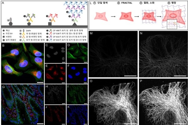
Through super-resolution microscopy (Stochastic Optical Reconstruction Microscopy) analysis, the team found out that antibodies formed uniform binding layers according to the number of staining and that they amplified fluorescence signals. It implemented simultaneous multi-label signal amplification imaging and signal amplification technology with high fluorescence intensity even after expansion when the technique was also applied to an expansion microscope (microscope that obtains superhigh resolution by expanding tissue)
This technique is expected to contribute towards analysis of body tissues through imaging and development of necessary treatments, multivariate index assay, and medical and new drug development.
“This technique that has a high level of image processing will play a vital role in developing the digital pathology field.” said Cho Ye-rin who is the first author and a student at the KAIST’s Department of Materials Science and Engineering. “Because it provides accurate information about multivariate index within the human body, it can be directly applied to analysis of medicine and medical supplies and treatment systems in the modern medical field.”
“While the technique becomes very important within the inside of a tissue during biopsy on a patient, it can also accurately image biomarkers that are revealed at a very low level.” said Professor Jang. “We expect that thee technique will be a great help in cancer diagnosis and estimating reaction rate of anticancer drugs.”
Staff Reporter Kim, Youngjoon | kyj85@etnews.com
Results on multilabel imaging and expansion microscopy when FRACTAL technique was applied